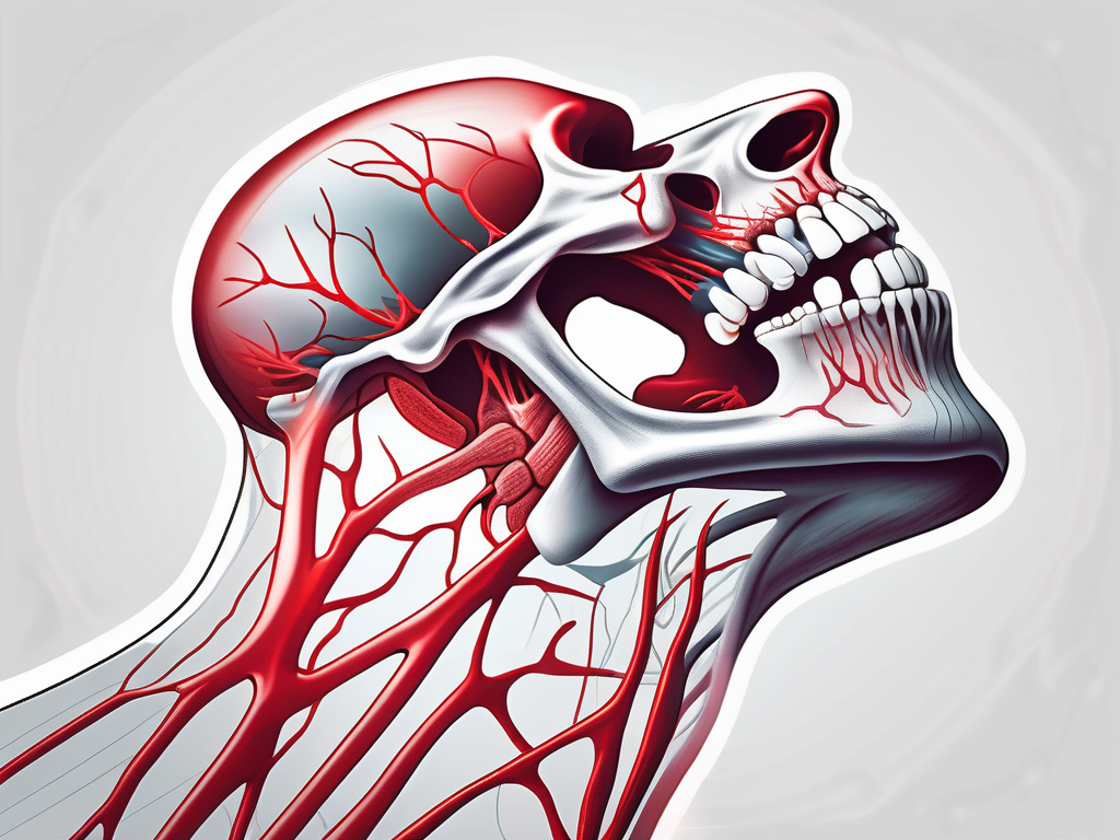
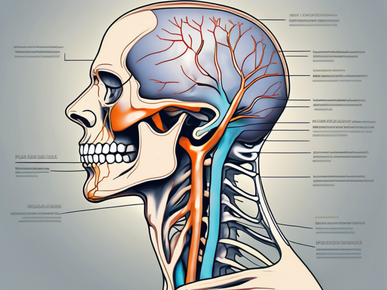
why does my mandibular nerve hurt
Read More
17 mins read

Discover the fascinating world of the mandibular nerve and its extensive innervations.

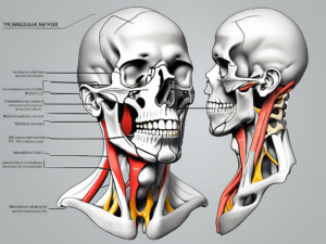
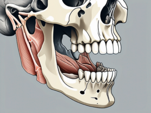
Discover the intricate relationship between the mandibular nerve and the muscles it innervates in this comprehensive article.
Discover the intricate functions and significance of the mandibular nerve in our latest article.

Discover the role and significance of the mandibular nerve, a division of the trigeminal nerve, in this comprehensive article.
Learn effective and quick techniques to heal the mandibular nerve with our comprehensive guide.
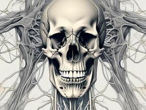
Discover the intricate network of nerves in the human body as you explore what the mandibular nerve innervates.
Discover the ins and outs of performing a mandibular nerve block in this comprehensive article.
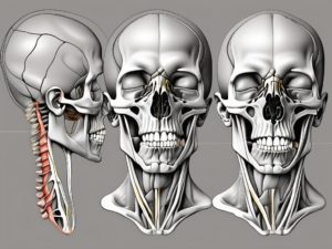
Discover the intricate network of the mandibular nerve as it intricately innervates the mylohyoid muscle.
Discover the fascinating world of the mandibular nerve and uncover the intricate processes that occur when it is moved.

Experience a jolt of discomfort? Discover the reasons why your mandibular nerve flares up when you drink coffee and learn how to ease the discomfort.