what does the mandibular nerve pass through dog skull
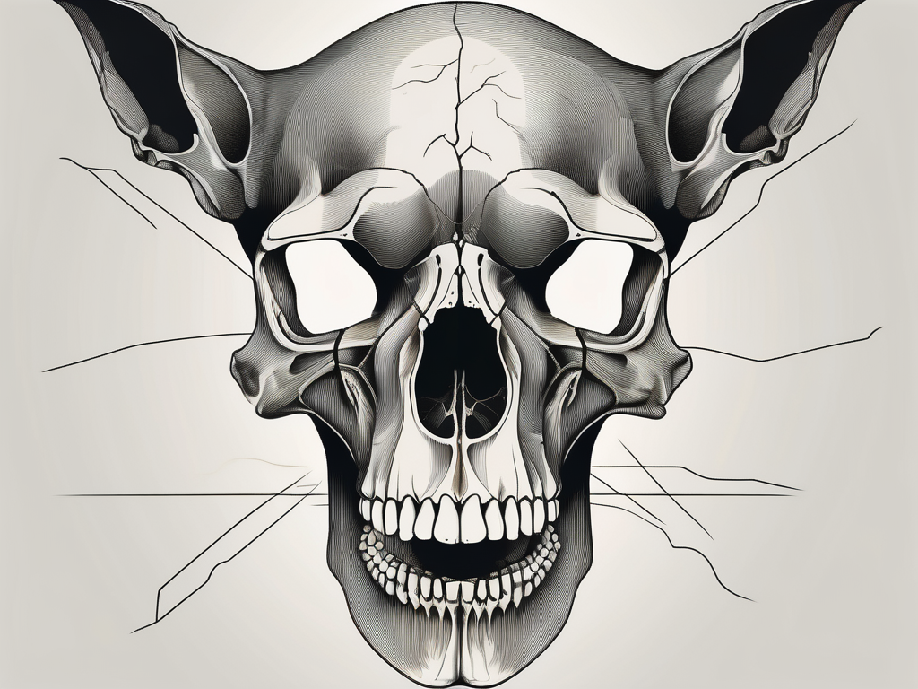
The mandibular nerve is an essential component of a dog’s anatomy, playing a vital role in the overall functionality of the animal’s skull. Understanding the intricate relationship between the mandibular nerve and the dog skull is crucial for veterinarians and pet owners alike, as it provides insight into potential problems and disorders that may arise.
Understanding the Mandibular Nerve in Dogs
The mandibular nerve is one of the three branches of the trigeminal nerve, which is responsible for providing sensory innervation to the face and motor control to the muscles involved in chewing. In dogs, the mandibular nerve is significant as it traverses through various structures within the skull, allowing it to carry out its critical functions.
Anatomy of the Mandibular Nerve
The mandibular nerve originates from the trigeminal ganglion, located within the skull. It descends from its point of origin, passing through the foramen ovale, a bony aperture in the base of the skull. As it exits the skull, the mandibular nerve branches out into smaller nerves, supplying sensation to the lower jaw, teeth, and other facial structures.
Within the skull, the mandibular nerve travels alongside other important structures, such as the maxillary nerve and ophthalmic nerve. These nerves work together to ensure proper sensory and motor function of the face. The intricate network of nerves within the skull allows for precise coordination and control of facial movements.
As the mandibular nerve continues its course, it gives off branches that innervate specific regions of the face. One such branch is the buccal nerve, which provides sensation to the cheek and the skin around the mouth. Another branch, the auriculotemporal nerve, supplies sensation to the external ear and the temporal region of the head.
Function of the Mandibular Nerve
The primary function of the mandibular nerve is to provide sensory information and innervation to various parts of a dog’s face. This includes the skin on the lower jaw, gums, teeth, and tongue. Additionally, it controls the muscles of mastication, allowing for efficient chewing and grinding of food.
The mandibular nerve plays a crucial role in a dog’s ability to eat and drink. It provides the necessary sensory feedback to ensure proper placement and movement of the jaw during chewing. Without the mandibular nerve, a dog would struggle to chew food effectively, leading to difficulties in obtaining proper nutrition.
Furthermore, the mandibular nerve is involved in the regulation of facial expressions. It controls the muscles responsible for various facial movements, such as opening and closing the mouth, raising the lips, and wrinkling the nose. These movements are essential for communication and expressing emotions.
It is important to note that any disruption or damage to the mandibular nerve can result in significant functional impairments, affecting a dog’s ability to eat, drink, and display normal facial movements. Therefore, an in-depth understanding of the mandibular nerve’s role is invaluable in diagnosing and treating potential problems.
The Dog Skull: An Overview
The dog skull is a remarkable structure, encompassing various interconnected bone segments that provide protection to the brain and support to the facial structures. Understanding the basic structure and key features of a dog’s skull is essential to comprehend the path of the mandibular nerve through this intricate framework.
When examining a dog’s skull, it becomes evident that it is a complex arrangement of bones, each with its own unique purpose. The cranium, for example, forms the protective casing around the brain, shielding it from potential injuries. The mandible, on the other hand, is responsible for housing the lower teeth and providing support for the lower jaw. The maxilla, zygomatic arch, and nasal bones all work together to form the upper jaw and contribute to the overall structure and function of the skull.
Basic Structure of a Dog’s Skull
A dog’s skull is composed of numerous bones that articulate with each other through specialized joints called sutures. These sutures allow for slight movement and flexibility, which is crucial for the dog’s ability to chew, bite, and express various facial expressions. The intricate network of bones ensures that the skull remains strong and stable, while still allowing for necessary mobility.
One fascinating aspect of a dog’s skull is the presence of fontanelles, which are soft spots found in the skull of puppies. These fontanelles allow for the growth and expansion of the skull as the puppy develops, gradually closing as the bones fuse together. This process ensures that the skull reaches its full strength and stability as the dog matures.
Key Features of a Dog’s Skull
Several key features of a dog’s skull contribute to its unique characteristics. One such feature is the presence of a well-defined stop, which is the abrupt transition between the forehead and muzzle. This stop gives dogs their distinctive facial appearance and varies in prominence depending on the breed. Additionally, the shape and arrangement of teeth within the mouth are essential features to consider when examining a dog’s skull.
The teeth of a dog serve various functions, including tearing, cutting, and grinding food. The arrangement of these teeth, such as the presence of canines for gripping and tearing, molars for grinding, and incisors for biting, reflects the dog’s dietary habits and evolutionary adaptations. By examining the teeth within a dog’s skull, veterinarians can gain valuable insights into the animal’s feeding behavior and overall health.
Understanding these features helps veterinarians assess the alignment of the mandibular nerve with the rest of the skull. The mandibular nerve is a crucial component of the dog’s nervous system, responsible for transmitting sensory information from the lower jaw and controlling the movement of the muscles involved in chewing. By comprehending the intricate relationship between the mandibular nerve and the dog’s skull, veterinarians can diagnose and treat various dental and neurological conditions more effectively.
The Path of the Mandibular Nerve Through the Dog Skull
The mandibular nerve follows a distinct path as it traverses through the dog skull. This path is characterized by its origin, route, and termination, each playing a crucial role in the nerve’s functionality and the overall health of the dog.
Understanding the intricate details of the mandibular nerve’s journey can provide valuable insights into the complex network of nerves that contribute to a dog’s ability to eat, chew, and perceive sensations in the lower part of its face.
Origin of the Mandibular Nerve
As previously mentioned, the mandibular nerve originates from the trigeminal ganglion, a cluster of nerve cell bodies located within the skull. This ganglion serves as a central hub for various sensory nerves, including the mandibular nerve.
From its origin, the mandibular nerve descends towards the foramen ovale, a bony aperture located in the skull. This opening acts as a gateway for the nerve, allowing it to exit the protective confines of the skull and venture into the surrounding structures.
Route of the Mandibular Nerve
Exiting the skull through the foramen ovale, the mandibular nerve embarks on a complex course, navigating its way through the intricate anatomy of the dog’s head. Along its route, the nerve gives rise to numerous branches, each with a specific function and destination.
One of the primary branches of the mandibular nerve is responsible for innervating the lower jaw, providing the necessary sensory input for the dog’s ability to chew, bite, and manipulate food. This branch sends signals to the muscles involved in chewing, ensuring the coordination and strength required for effective mastication.
In addition to its role in jaw movement, the mandibular nerve also supplies sensory fibers to the teeth, gums, and tongue. These fibers enable the dog to perceive tactile sensations, such as pressure and texture, allowing for the detection of potential dental issues or foreign objects in the mouth.
Termination of the Mandibular Nerve
The mandibular nerve reaches its termination point in the lower jaw region, where its sensory fibers reach the skin and mucous membranes. This final destination is crucial for the dog’s ability to perceive touch, pain, and temperature sensations in the lower part of its face.
By terminating in the lower jaw region, the mandibular nerve ensures that the dog can accurately sense and respond to external stimuli. This includes the ability to detect changes in temperature, which can help the dog avoid potential harm or discomfort.
Furthermore, the termination of the mandibular nerve in the lower jaw region allows for the perception of pain, serving as a warning mechanism in case of injury or inflammation. This sensory feedback plays a vital role in the dog’s overall well-being, as it enables the identification and prompt response to potential health issues.
In conclusion, the path of the mandibular nerve through the dog skull is a complex and intricate journey. From its origin in the trigeminal ganglion to its termination in the lower jaw region, this nerve plays a crucial role in the dog’s ability to eat, chew, and perceive sensory information in the lower part of its face. Understanding the details of this path can provide valuable insights into the intricate workings of a dog’s nervous system and its overall health.
The Relationship Between the Mandibular Nerve and Dog Skull
The intricate relationship between the mandibular nerve and the dog skull is of paramount importance, as any disruptions to this relationship can have significant implications for a dog’s health and well-being.
How the Mandibular Nerve Interacts with the Skull
The mandibular nerve interacts with the skull through its passage within the foramen ovale. This bony aperture provides a protective conduit, ensuring the nerve remains secure during its journey. The foramen ovale, located on the ventral surface of the skull, is a crucial anatomical feature that allows the mandibular nerve to exit the cranial cavity and enter the mandibular region.
As the mandibular nerve traverses the foramen ovale, it is surrounded by various structures, including blood vessels, connective tissues, and other nerves. These neighboring structures contribute to the overall stability and support of the mandibular nerve, preventing it from being easily compressed or damaged. The intricate arrangement of these structures within the skull showcases the remarkable design of the canine anatomy.
Any compromise to the integrity of the foramen ovale or other skull structures can result in compression or damage to the mandibular nerve, leading to functional deficits. Trauma, such as fractures or dislocations of the skull, can disrupt the normal relationship between the mandibular nerve and the skull. Additionally, certain pathological conditions, such as tumors or infections, can also affect the mandibular nerve’s interaction with the skull, further highlighting the delicate nature of this relationship.
Importance of the Mandibular Nerve’s Path
The path of the mandibular nerve is critical to maintaining proper sensory and motor function in a dog’s face. As the mandibular nerve branches out from the skull, it innervates various structures, including the lower jaw, lower lip, and the muscles responsible for chewing. This extensive innervation allows dogs to perform essential functions such as eating, grooming, and expressing facial expressions.
Disruption of the mandibular nerve’s path, whether due to trauma, disease, or anatomical abnormalities, can result in conditions such as facial paralysis, difficulty eating, and compromised dental health. Facial paralysis can lead to drooping of the lower lip, asymmetrical facial expressions, and difficulty closing the mouth properly. Additionally, compromised sensory function in the lower jaw can affect a dog’s ability to detect pain, temperature, and pressure, potentially leading to oral health issues that may go unnoticed.
Recognizing and addressing issues related to the mandibular nerve requires a comprehensive understanding of its route through the skull. Veterinarians and veterinary specialists utilize advanced imaging techniques, such as computed tomography (CT) scans or magnetic resonance imaging (MRI), to visualize the mandibular nerve’s pathway and identify any abnormalities or potential sources of compression. This detailed knowledge allows for accurate diagnosis and targeted treatment, ensuring the best possible outcome for dogs experiencing issues related to the mandibular nerve and skull relationship.
Potential Problems and Disorders
While the mandibular nerve is vital for a dog’s overall health, various problems and disorders can affect its functionality. Recognizing the signs and symptoms of these conditions is crucial for early detection and appropriate intervention.
The mandibular nerve is responsible for innervating the muscles of mastication, allowing dogs to chew and eat their food. It also provides sensation to the lower jaw, lower lip, and part of the tongue. Any disruption in its normal function can lead to discomfort and difficulty in performing these essential activities.
Common Disorders Affecting the Mandibular Nerve
Disorders commonly affecting the mandibular nerve include nerve entrapment, inflammation, infection, and neoplasia. Nerve entrapment occurs when the nerve becomes compressed or trapped, leading to impaired nerve conduction and subsequent symptoms. Inflammation can result from various causes, such as trauma or autoimmune conditions, and can lead to pain and dysfunction. Infections, including abscesses or dental infections, can also affect the mandibular nerve, causing localized swelling and discomfort. Neoplasia, or the presence of tumors, can disrupt the normal function of the nerve and may require more extensive treatment.
Each of these conditions can result in pain, numbness, weakness, or loss of sensation in the areas innervated by the mandibular nerve. Dogs may experience difficulty in chewing or opening and closing their mouths properly. Additionally, dental diseases, such as periodontal disease or fractured teeth, can also impact the mandibular nerve’s functions.
Symptoms and Diagnosis of Mandibular Nerve Disorders
Recognizing the symptoms associated with mandibular nerve disorders is paramount for diagnosis and appropriate management. Owners should be vigilant for any changes in their dog’s behavior or physical appearance that may indicate a problem. Symptoms may include drooping of the lower jaw, difficulty in opening or closing the mouth, changes in chewing behavior, facial swelling, and oral sensitivity.
If your dog is experiencing any of these symptoms, it is important to consult with a veterinarian who can provide an accurate diagnosis and recommend the most suitable treatment options. A thorough physical examination will be conducted, focusing on the jaw and oral cavity. Diagnostic imaging techniques, such as radiographs or CT scans, may be necessary to identify the underlying cause and assess the extent of the damage.
Furthermore, a comprehensive dental examination will be performed to evaluate the oral health of your dog. This may involve a thorough cleaning, dental X-rays, and assessment of any dental abnormalities or pathology that may be contributing to the mandibular nerve disorder.
Treatment Options for Mandibular Nerve Disorders
The treatment for mandibular nerve disorders depends on the specific cause and severity of the condition. In some cases, conservative management, including pain management and anti-inflammatory medications, may be sufficient to alleviate symptoms and restore normal function. Physical therapy and rehabilitation exercises may also be recommended to improve muscle strength and coordination.
However, more complex cases may require surgical intervention or specialized therapies to address underlying pathology. Nerve decompression surgery may be performed to relieve pressure on the nerve and restore its function. In cases of infection or neoplasia, appropriate treatment, such as antibiotics or tumor removal, will be necessary to eliminate the source of the problem.
It is essential to consult with your veterinarian to determine the best course of action for your dog. Professional guidance and expertise will ensure the appropriate treatment plan is developed and implemented. Your veterinarian will consider factors such as your dog’s overall health, the severity of the condition, and your preferences as the owner when recommending the most suitable treatment options.
Remember, early detection and intervention are key to successful outcomes in managing mandibular nerve disorders. By staying vigilant and seeking prompt veterinary care, you can help ensure your dog’s comfort and quality of life.
Conclusion: The Mandibular Nerve’s Role in a Dog’s Health
The mandibular nerve’s path through the dog skull is a fascinating and complex journey that plays a critical role in a dog’s health and well-being. Understanding the anatomy, function, and potential disorders associated with this nerve provides invaluable insights for veterinarians and pet owners alike.
Should you notice any concerning symptoms related to your dog’s jaw, mouth, or facial functions, it is essential to seek veterinary attention promptly. Only a trained professional can accurately diagnose and treat any potential mandibular nerve disorders, ensuring your furry companion receives the care and support they need for a healthy and fulfilling life.


